 Aim
Aim
Becoming familiar with compound microscope ; discovering the biological world of small and minute
Class level : 9th-12th grade;13 – 18 year olds.
Introduction:
This activity is aimed at making you become familiar with compound microscope and be able to use it with ease. A whole world opens up to you when you learn to use the microscope
Materials needed :
Glass slides, cover slips, petri dish/ watch glass, chemical stains, dropper, mounted needle, glycerine, a thin brush and water in a beaker.
Biological specimens needed: You can use any one of the following, but I would suggest that you start by looking at the pollen grains.Other materials that can be used : Onion peel, Epidermal leaf peel which you can obtain from monocot leaves that are thick and juicy; spores of fungi; cotton, woolen or silk yarn; sand grains.
A word about pollen grains :
Pollen grains of plants are unique to each species. Species can be identified by looking at their pollen grains because of the differences. What is different? Can you find out? Try this activity.
Warning : MANY ARE ALLERGIC TO POLLENS. CONSULT YOUR TEACHER/PARENTS BEFORE ATTEMPTING THIS ACTIVITY.
Observing pollens under the microscope : Suggested plants : Spider lilly, commonly seen Hibiscus( the Red variety),Crotolaria sp; Commelina sp., Tradescantia etc.or choose one which is available in your neighbourhood. Choose flowers in which anthers are dehiscing.
Step 1 :
Hibiscus : Choose the flower for your activity carefully. If you observe you will see that the anthers are (dehiscing) splitting and the pollen is ready for release. Choose such flowers.
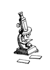
Step 2 : Set your microscope ready ( as described earlier) on the table.
Step 3 : Gently hold the stamen by the filament (close to the anther) and tap the anther on to the glass slide. You will see yellow powder on your glass slide. Add a drop of water and place the slide under the 10x objective lens for viewing. Looking through the eye piece raise the body tube up so as to bring the material into focus. What do you see?
- Note down your observations. Draw the pollen grain.
Step 4 :If you wish to see more details then use the 45x objective lens. But for this type of viewing you will have to first cover the pollen grains with a cover slip. If you do not do this then the objective lens would be spoiled as it would then come in contact with water from your slide.Be careful while using the higher magnification as the objective lens will have to be brought very close to the slide to focus. For fine focusing use the fine adjustment screw and not the coarse adjustment one.
Under higher magnification you would be able to see the spiny outer covering of the pollen grain. This is called exine. If you are lucky you may even see a germinating pollen grain, where the exine has split and the pollen tube is beginning to emerge.

Pollen Grains

Exine
See also http://geography.berkeley.edu/ProjectsResources/PollenKey/byFamiliesAll-in-1.html#Compositae
This website gives you micrographs of pollens of plants from different families. Though these are not plants from India, it will never the less give you an idea of what you are able to see under the microscope
Repeat steps 1- 3 for viewing pollen grains from other flowers. Are the shapes,sizes, designs on pollens from different plants the same? You can actually build up a data base on pollens of the plants from your area.
Extension of this activity:
Should you see a pollen with a pollen tube, then you can extend and modify this activity further.Take a watch glass and gently tap the anthers over its surface. When you see pollens, then add a drop of stain– either acetocarmine or any neutral red. After a minute, with the help of a brush transfer the pollen grains on to a glass slide. Add a drop of glycerine ( so that the pollen tube does not dry up ) and place a cover slip gently over it ensuring that there are no air bubbles. Place this slide under the low power of the microscope , focus and observe.
- Note down your observations. Draw what you observe.
Gently move the nose piece and align the higher objective lens ( 45x or 60x) above the slide. Focus and observe.
- Note down your observations. What are the details that are now visible? Draw and mark the parts you observe.
NOTE :
- Sometimes pollens can be found in dehydrated form. If you see shriveled up forms under the microscope, then prepare the slide again. First dust the anthers on the watch glass and add water. Leave them for some time to absorb water and become hydrated.
- Ensure that the pollens you are studying is of that flower and has not been contaminated by pollen grains from other plants. For eg wind may have blown some pollen grains on to this flower.
Observing cotton fibres :
A variety of plant fibres may be observed under the microscope. Take some cotton from the cotton plant ( or else from the cotton from the first-aid box). Separate a thin fibre place it on the slide, set your microscope and observe. Play around seeing it under different magnification. What do you observe?
Observing the butterfly wing scales
Butterfly scales are fascinating to see under the microscope. Do not catch a live butterfly if possible. Ideally you should get a dead portion of the butterfly wing. Or else hold the butterfly gently ( GENTLY PLEASE) by its two wings (just to ensure that we do not harm the butterfly in any way we hold its two wings together) for a minute or so and then release it. Observe your fingers
You will see some powdery stuff left on your fingers. Gently transfer this powder to a glass slide and observe it using the 10x objective lens. Cover it with a cover slip and observe it under higher magnification. What do you observe?
If you are a naturalist and an outdoor portion then you will often see wings of insects among the grasses. If you see any collect them and see them under the microscope.
The microscopic world is for you to discover. Learn to design your own activities with the microscope. You will find it learning biology a great fun.
So what have you learnt from these activities?
Test yourself.
1. Given below are a few items (A)scientists and researchers use in microbiology laboratories. Match these items correctly with the activities (B) for which the researchers use them. Sometimes more than one of the items would be used for an activity
A – List of Items : Cover slips; Iodine stain; glycerine; water; 60x objective lens; Petridish; Methylene blue; Forceps; brush; watch glass.
B – Activities of the scientists and researchers –
- Culturing (growing)fungi that commonly grow on food items.
- Prepare slides for viewing under high power of microscope.
- Getting the microscope ready to view objects under higher magnification
- Observing to see whether carbohydrates are present.
- Preparing a slide of the human cheek cell to observe nucleus
- Ensuring that the prepared slide does not dry up fast.
- Hydrating the pollen grains before mounting them on a slide.
- Staining the human cheek cell to observe nucleus
- Separating the fibres or cotton/wool for preparing a slide
- Staining a material before placing it on a slide.
C – Give reasons for the following :
- Coverslip must be place over a slide before viewing it under 45x
- Glycerine is used to make a slide if it has to be used for a long period of study
- If 45x objective lens is not available then an 15 x eyepiece lens is sometimes used.
- Fine adjustment screw is used to get the image into focus when using the microscope for obtaining higher magnification


Leave a Reply